


Tired of Hedging in Your Pathology Reports? Here's What to Do About It
If your pathology reports frequently say 'consistent with' or 'cannot exclude,' the tissue isn't the problem. The context is. Here's what to include to get a more definitive answer.
Canine Mast Cell Tumor Grading: Understanding the Two Systems and What Your Report Is Actually Telling You
Canine MCT grading has improved significantly since 1984. It is still imperfect. Grade I MCTs can metastasize. Kiupel low-grade tumors can behave aggressively. A plain-language breakdown of both systems — and where AI grading might take us next.

Proteomics in Veterinary Oncology: A Deeper Look at What Tumors Are Actually Doing
Genomics tells us what mutations a tumor carries. Proteomics tells us what the tumor is actually doing with them. A look at where proteomics stands in veterinary oncology today — and where it's going.

Special Stains vs. Immunohistochemistry: What They Are and When Your Pathologist Uses Each
When your pathology report recommends additional testing, it's either chemistry or antibodies — and the choice between them tells you a lot about what question the case is raising.

Acanthomatous Ameloblastoma: When the Margins Tell the Real Story
Canine acanthomatous ameloblastoma doesn't metastasize. The entire prognosis lives in the surgical margin — and this case shows exactly why that number needs context to mean anything.
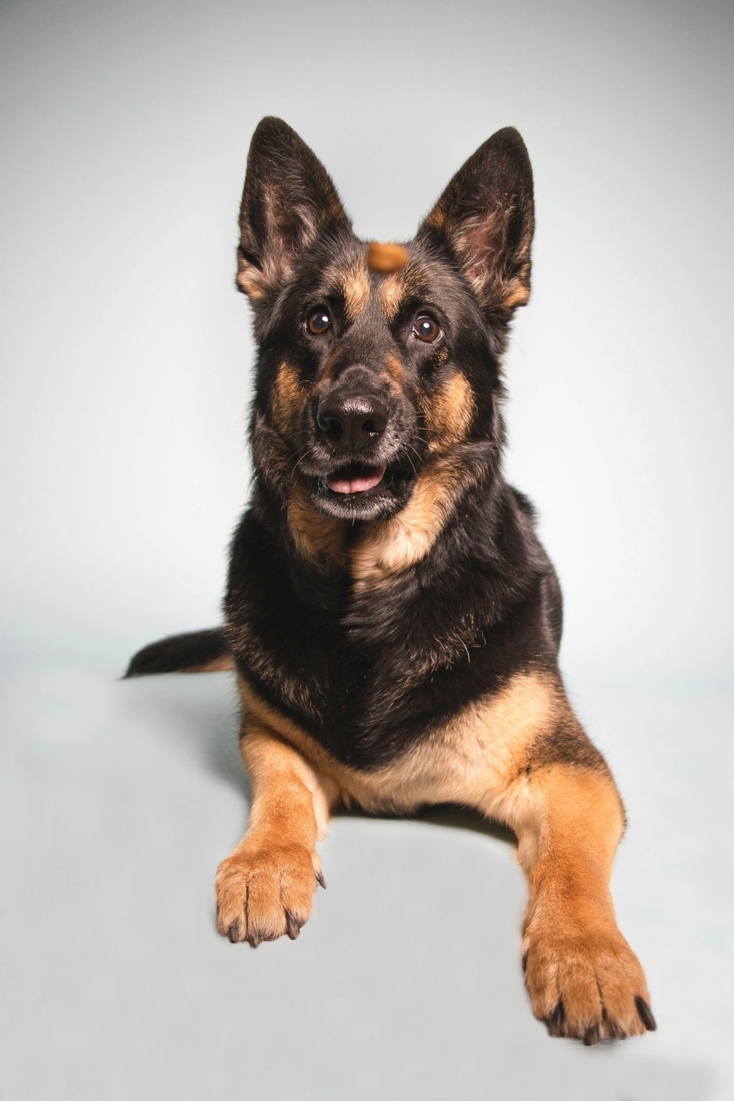
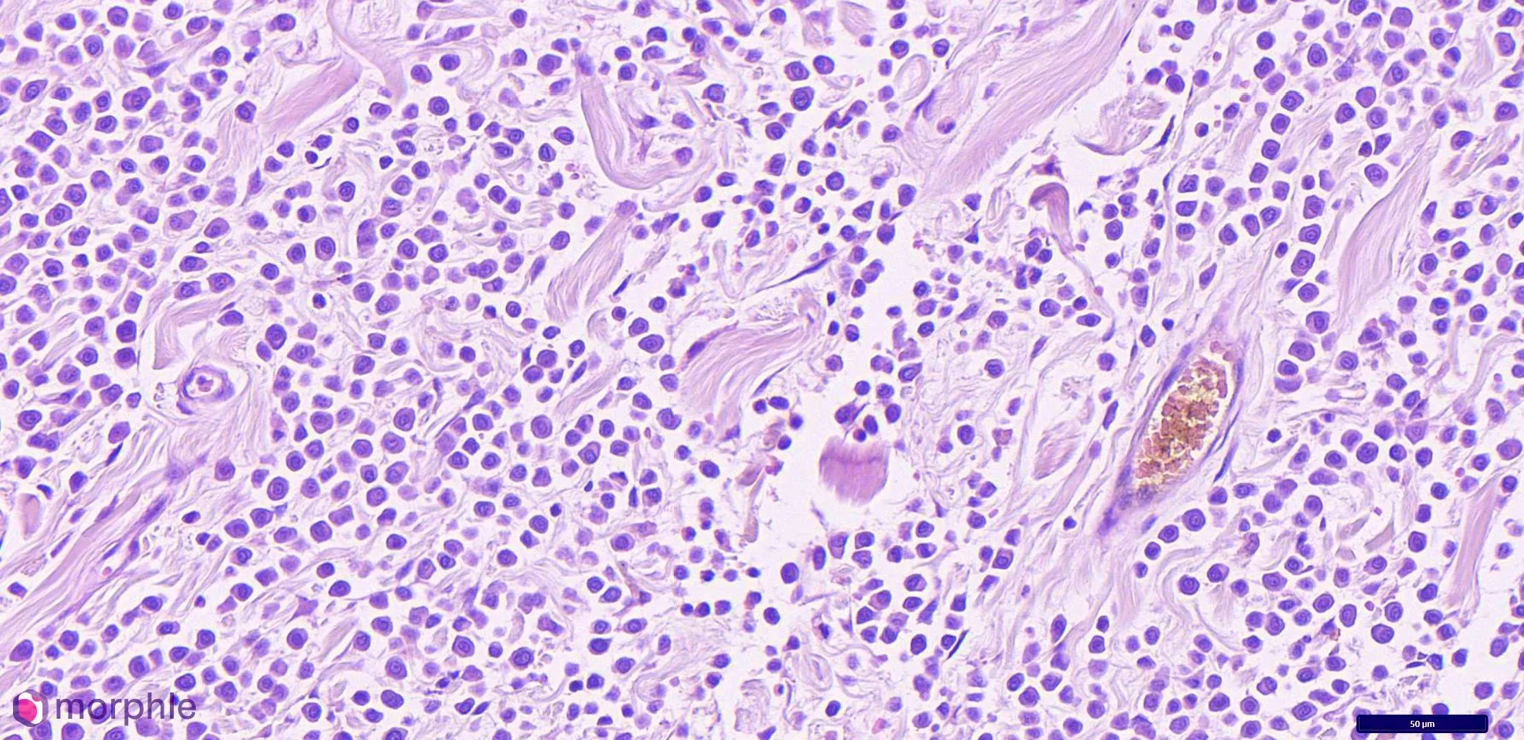
Nodular Dermatofibrosis and Renal Cystadenocarcinoma: Recognizing a Skin Lesion That Points Inward
Multiple firm nodules on a German Shepherd's legs have a differential list. Nodular dermatofibrosis should be on it — because the diagnosis it points to is bilateral renal cystadenocarcinoma

Liquid Biopsy and Circulating Tumor DNA: Where Veterinary Oncology Diagnostics Are Headed
Liquid biopsy is here. The question is no longer whether it has a role in veterinary oncology — it's understanding what that role is today and where it's going in the next few years.
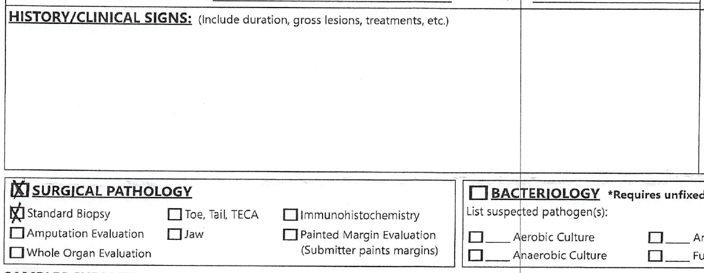
Necropsy Submissions: How to Maximize Diagnostic Yield
A step-by-step guide for veterinarians performing necropsies: how to handle carcasses, select and fix tissue, collect ancillary samples, and document findings so the histopathology report is as complete as possible
Feline Oral Squamous Cell Carcinoma: Why the Prognosis Is So Poor and What Histopathology Actually Contributes
FOSCC accounts for 60–70% of feline oral tumors and carries a uniformly poor prognosis. A board-certified veterinary pathologist explains the histologic features, bone invasion biology, and what a complete pathology report should communicate to your surgical team.
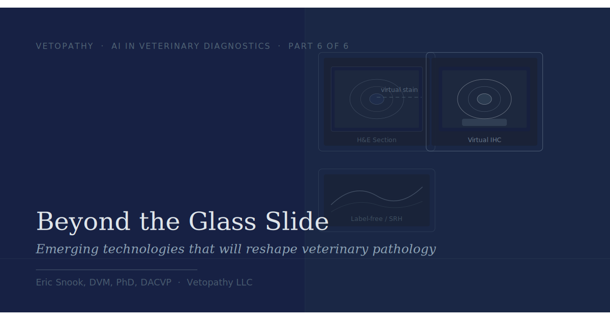
Beyond the Glass Slide: Emerging Technologies That Will Reshape Veterinary Pathology
The histopathology workflow has been essentially unchanged for decades. Two emerging technologies — virtual staining and label-free imaging — represent something genuinely different. Neither is ready for routine veterinary use yet. Both are worth understanding now.
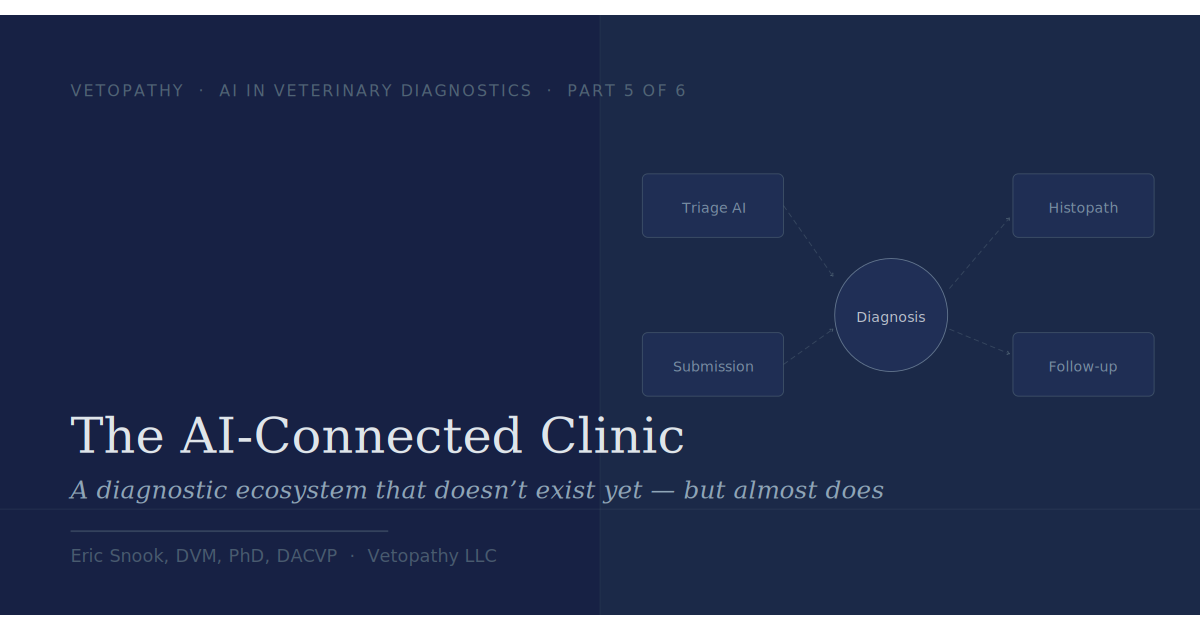
The AI-Connected Clinic: A Diagnostic Ecosystem That Doesn’t Exist Yet — But Almost Does
The individual AI tools reshaping veterinary diagnostics each solve a piece of the puzzle. The bigger opportunity — and the harder one — is connecting them. Here's what that could look like, why it matters, and what practices can do right now to build toward it.
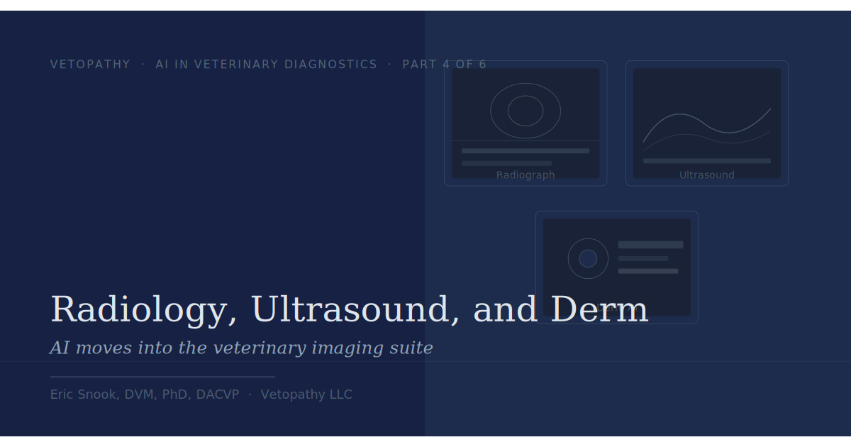
Radiology, Ultrasound, and Derm: AI Moves Into the Veterinary Imaging Suite
Imaging data has properties that make it a natural fit for AI. It's inherently digital, produced in large volumes, and involves the kind of spatial pattern recognition that machine learning handles well. This is why medical imaging was one of the first clinical areas where AI showed real promise — and why veterinary imaging is following a similar path, with a lag that reflects the smaller scale of the veterinary market rather than any fundamental barrier.
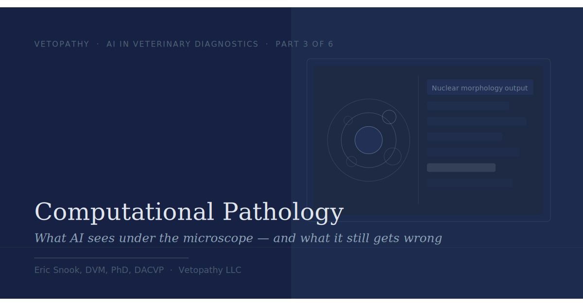
Computational Pathology: What AI Sees Under the Microscope — and What It Still Gets Wrong
Computational pathology is the application of digital image analysis and machine learning to tissue and cytology samples. It covers a wide range of tasks that differ significantly in how technically complex they are and how well they've been validated.
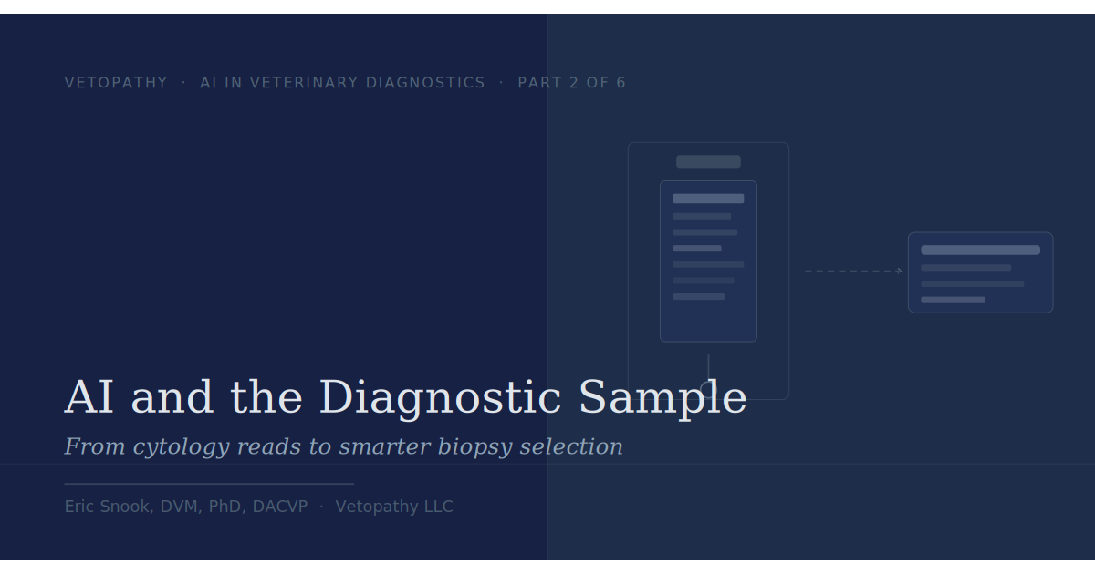
AI and the Diagnostic Sample: From Cytology Reads to Smarter Biopsy Selection
The diagnostic sample is where clinical impressions become something testable — and where a surprising amount of diagnostic information is lost before it ever reaches the lab. AI is beginning to change what gets sampled, how it's documented, and whether it gets submitted at all.
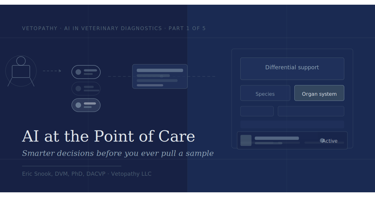
AI at the Point of Care
That is where artificial intelligence stands the most immediate chance of making a difference in veterinary medicine. Not in replacing the diagnostic expertise at the end of the pipeline, but in improving the quality of what enters it.

Beyond Lymphocytic-Plasmacytic IBD: Recognizing Eosinophilic Enteritis at Surgery
Sections of small intestinal wall revealed a marked eosinophilic infiltrate throughout the lamina propria. Eosinophils were present in high numbers in all sections examined, with infiltration extending into the crypts and disrupting the normal crypt architecture in areas. Villous blunting and crypt irregularity were present, consistent with chronic mucosal injury. The degree of eosinophilic infiltration was inconsistent with a reactive response to the foreign body alone — the distribution and severity indicated a pre-existing, active inflammatory process.

Encephalitozoon cuniculi in Rabbits: A Pathogen Your Patients Are Likely Already Carrying
E. cuniculi sits in a diagnostically uncomfortable position: it is common enough to be a reflex differential in almost any sick rabbit, but the available antemortem diagnostics are imprecise enough that a positive titer is often adjunctive rather than confirmatory. Understanding the pathology underlying clinical presentations — and what histopathology can and cannot contribute — is essential for navigating these cases well.

Flow Cytometry for Lymphoma Immunophenotyping in Dogs and Cats: An Underutilized Tool
…flow cytometry remains underutilized in general practice. The purpose of this post is to explain what it does, what it does not do, and when it should be part of your diagnostic plan.
Mammary Carcinoma in a Male Cat: A Diagnosis Worth Not Missing
Male cats can develop mammary carcinoma. The diagnosis should be on the differential list for any mammary region mass in a male cat, regardless of neuter status. Given that the vast majority of feline mammary tumors are malignant, histopathology is not optional — cytology alone is unreliable for distinguishing benign from malignant mammary lesions in cats.