

Mammary Carcinoma in a Male Cat: A Diagnosis Worth Not Missing
Mammary neoplasia in cats is well-recognized in intact and ovariectomized females. In male cats, it is rare enough that the diagnosis can catch clinicians off guard — and that unfamiliarity is precisely why this case is worth documenting.
Signalment and Clinical Presentation
A 15-year-old neutered male domestic shorthair presented with spherical mass containing cystic and solid areas. The clinical impression at the time of submission was open. An inverted nipple had previously been identified in October 2025. For any number of reasons, mammary carcinoma was not the top consideration of the clinician at the time of excision.
The mass was submitted for histopathology following excision.
Histopathologic Findings
Sections revealed a malignant epithelial neoplasm with an infiltrative growth pattern. Neoplastic cells formed irregular tubular and solid nests separated by a moderate fibrous stroma. The infiltrative margin was a prominent feature — neoplastic cells extended into and through the surrounding soft tissue without a well-defined capsule.
The mitotic index was 11 per 2.37mm2 (not extraordinarily high); however, high enough that paired with the infiltrative growth pattern warrants a malignant diagnosis. Though, lymphovascular invasion was not identified, the neoplastic cells often encroached in vessels, which increases the suspicion for invasion in deeper sections.
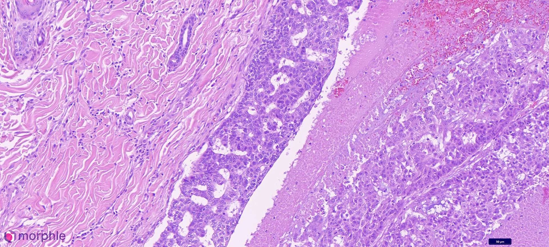
Mammary carcinoma in a male cat. This image demonstrates the edge of the neoplasm. The area to the right of center demonstrates an area of necrosis with viable epithelial cells. The area to the left is normal dermal stroma.
Diagnosis
Mammary carcinoma, infiltrative, low grade (according to Mills et al grading scheme).
The histologic features were consistent with malignant mammary origin. Because the cells were clearly epithelial in morphology, there was no need for immunohistochemical characterization of this neoplasm.
Discussion: Mammary Tumors in Male Cats
Mammary tumors in male cats are uncommon — estimated at roughly 1–2% of all feline mammary neoplasms. Because of this rarity, the published literature on prognosis and biologic behavior in male cats specifically is limited. What is established is that mammary tumors in cats as a species are predominantly malignant (approximately 85–90%), in contrast to the dog, where roughly half are benign. There is no compelling reason to expect male cats to differ meaningfully from this general pattern, and the present case is consistent with it.
Neuter status in males has not been shown to confer the same protective effect it does in females. In females, early ovariohysterectomy dramatically reduces mammary tumor risk; the hormonal dependency appears less straightforward in males, and most reported male cases are in neutered cats, as here.
The infiltrative growth pattern and high mitotic index in this case are histologic indicators of aggressive behavior. In female cats, high mitotic index and lack of circumscription are associated with shorter survival times and higher recurrence rates. By analogy, these findings in a male cat should be interpreted with the same prognostic weight.
The absence of lymph node submission is worth noting. In any mammary carcinoma — regardless of patient sex — regional lymph node evaluation allows for staging, which may help in clinical decision making. Axillary and inguinal lymph nodes are the primary regional targets depending on gland location.
Clinical Takeaway
Male cats can develop mammary carcinoma. The diagnosis should be on the differential list for any mammary region mass in a male cat, regardless of neuter status. Given that the vast majority of feline mammary tumors are malignant, histopathology is highly recommended — cytology alone is unreliable for distinguishing benign from malignant mammary lesions in cats.
When a mammary mass is identified in a male cat: excise with adequate margins, submit regional lymph nodes if feasible, and submit the mass for histopathology.
Case submitted to Vetopathy. Diagnosis rendered by Eric Snook, DVM, PhD, DACVP.